Here, I present my recent scientific work using volume imaging technology.
Dr. Csaba Adori
I am a neurobiologist currently working at Stockholm University, Department of Molecular Biosciences. I am also affiliated to the Department of Neuroscience at Karolinska Institutet, Stockholm, Sweden.
I have always been interested in how structure (on the macromolecular-, cellular-, histological- or on higher structural levels) determines functions and how impaired functionality is reflected in structural alterations.
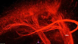
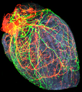
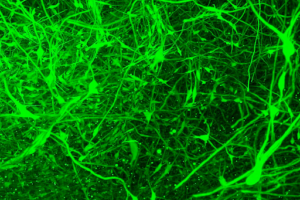
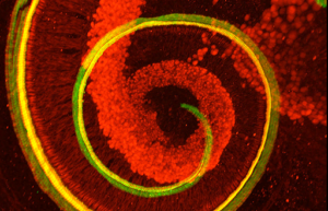
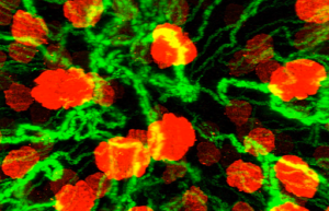
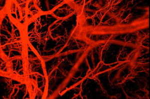
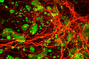
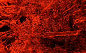
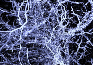
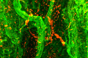
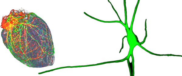
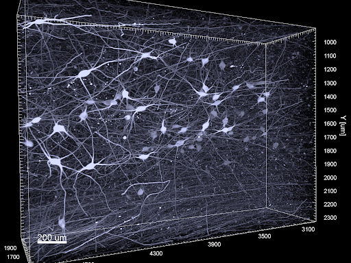
My particular research interest is about the anatomy and pathology of the serotonergic and noradrenergic systems. Some years ago, I started working with three-dimensional immuno-imaging and light sheet microscopy, which can provide lots of exciting details regarding the complex cytoarchitectural organization and projections of monoaminergic neurons in health and disease.
On this page, I introduce my recent work with three-dimensional imaging: first, my ongoing and completed scientific projects. In addition, I provide a systematic collection of short movies that demonstrate mammalian (including human) tissues in three dimensions, as we have never seen before.
The beauty of some of these pictures may reiterate the harmony of structure and function in nature and perhaps reminds us of the importance of artistic aspects in scientific cognition.

Dr. Csaba Adori
Researcher
Stockholm University
Department of Molecular Biosciences
Svante Arrhenius väg 20C
10691 Stockholm, Sweden

Karolinska Institutet
Department of Neuroscience
Biomedicum D7, Solnavägen 9,
17165 Solna

Email1: csaba.adori@ki.se
Email2: csaba.adori@su.se
Email3: adorics@gmail.com
Telephone: +46 7 09983554
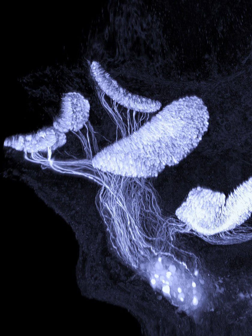
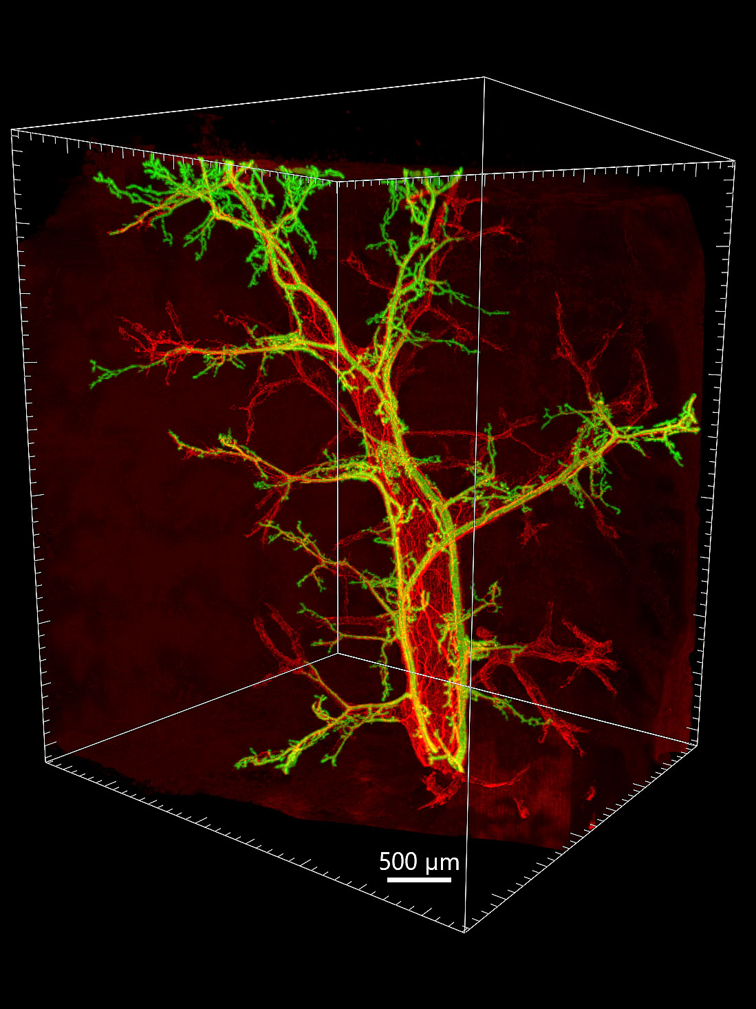
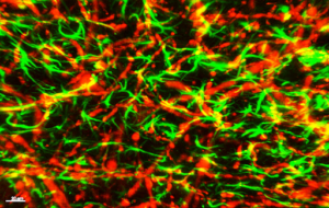
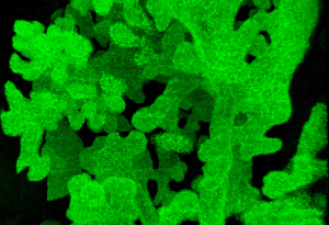
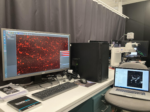
Staff portal at KI homepage STAFF PORTAL AT SU HOMEPAGEI use the iDISCO+ volume immunostaining and optical clearing technology. iDISCO means: immuno-enabled three-dimensional imaging of solvent cleared organs.
During the wet lab process, fixed tissue blocks, in my case typically with the size of 0.2 – 1 cm in all the three dimensions, are immunostained in toto, using primary antibodies and fluorochrome-conjugated secondary antibodies. The stained blocks are optically cleared (made to be transparent) with a series of organic solvents.
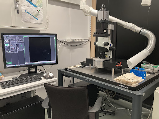
The stained and cleared blocks are scanned by a light sheet microscope (UM2, LaVision Biotec in our lab). Finally, the serials of images produced by the microscope are reconstructed in 3D by Imaris software (Bitplane). The reconstructed acquisitions can be used for comprehensive visual inspection and multiple 3D quantifications.
iDISCO.INFO HOMEPAGE FIRST DESCRIPTION OF iDISCO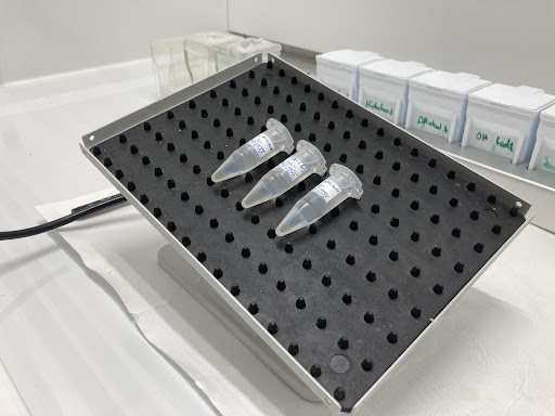
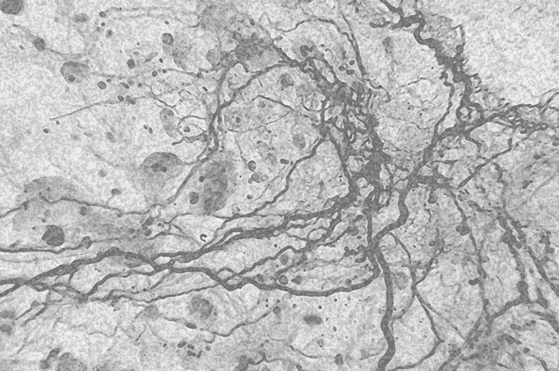
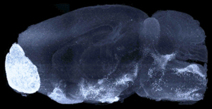
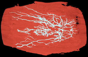
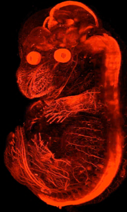
Here, I present several short movies about 3D structures of tissues which are shown in 18 different topics – including healthy and various pathologic conditions, in mouse and/or human tissues. All samples shown in these videos were fixed in 4% paraformaldehyde (modified Zamboni fixative).
By clicking on the icons, videos can be streamed on Youtube and YouKu and the higher resolution versions of the same videos can also be downloaded. In addition, brief descriptions are presented about each video, including the applied antibodies and tissues. For further information, please, contact: adorics@gmail.com.
If you use these videos in your presentation / publication, please cite this homepage accordingly: www.volume-
Many of these movies were used to create short educational films that are published in the Human Protein Atlas YouTube channel:
HPA video channel